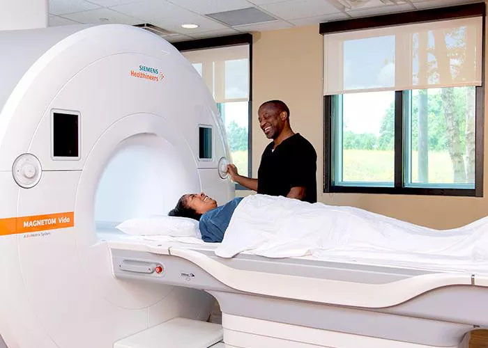
Magnetic Resonance Imaging (MRI) is one of the most advanced, non-invasive medical imaging techniques used to diagnose and monitor a wide range of health conditions. MRI machines have revolutionized the medical field by offering detailed images of the internal structures of the body, particularly the soft tissues. Unlike X-ray or CT scans, which use ionizing radiation, MRI relies on magnetic fields and radio waves to generate images, making it a safer option for patients, especially for those who need frequent imaging.
This article will delve into the working principles of MRI machines, their components, how they function, their uses, and their significance in modern medicine. By understanding the underlying science and mechanics of MRI machines, we can appreciate their crucial role in medical diagnostics.
What is MRI?
MRI stands for Magnetic Resonance Imaging, and it is a diagnostic technique that uses strong magnetic fields and radio waves to produce detailed images of the organs and tissues inside the body. These images can be used to detect abnormalities, diagnose diseases, or monitor the progression of various conditions.
Unlike traditional X-ray or CT scans, which use radiation, MRI relies on the principles of nuclear magnetic resonance (NMR). In NMR, atomic nuclei are exposed to a magnetic field and radiofrequency pulses, causing them to resonate and emit signals that are then captured and used to construct images. This technique can generate high-resolution images, particularly of soft tissues, like the brain, muscles, and organs, making it invaluable in various medical fields.
The Basic Components of an MRI Machine
To better understand how MRI machines work, it’s important to look at their key components. The basic structure of an MRI machine includes the following:
Magnet
The most critical part of an MRI machine is its powerful magnet, typically made from superconducting materials. The magnet creates a strong magnetic field, usually measured in Tesla (T), that aligns the hydrogen nuclei in the body. The typical strength of an MRI magnet is between 1.5 to 3 Tesla, though high-field MRI machines can go up to 7 Tesla for specialized applications.
Gradient Coils
Gradient coils are responsible for creating varying magnetic fields within the MRI machine. These coils adjust the magnetic field locally in specific areas to enable the precise location of signals within the body. By varying the magnetic field across space, the gradient coils allow the MRI machine to acquire images slice by slice, building a complete 3D image of the body part being scanned.
Radiofrequency (RF) Coils
RF coils are used to transmit and receive the radiofrequency signals required to produce the images. These coils emit radiofrequency pulses that excite hydrogen nuclei (protons) in the body. After the protons absorb the energy from the radiofrequency pulse, they resonate and emit signals, which the RF coils then detect.
Computer System and Image Processor
The computer system is the brain of the MRI machine. After the RF coils capture the emitted signals from the protons, the computer processes this data and translates it into images. This step involves complex algorithms that reconstruct the data into two-dimensional or three-dimensional images of the body’s internal structures.
Patient Table
The patient lies on a movable table that is positioned inside the MRI machine. The table can slide into the magnet, allowing the body part being examined to be exposed to the magnetic field. For certain MRI procedures, the table can move to adjust the area of interest in the scanner.
Shielding and Safety Mechanisms
Since MRI machines use strong magnetic fields, they are typically housed in a well-shielded room to prevent interference with external devices and to protect people from magnetic exposure. The room often contains metal shielding, and safety procedures ensure that ferromagnetic objects are kept away from the machine.
How MRI Machines Work: The Underlying Science
The mechanism behind MRI involves the interaction between the magnetic field and hydrogen nuclei (protons) found abundantly in the body, particularly in water molecules. Here’s a step-by-step explanation of how MRI works:
Aligning the Protons
The body is made up of about 60-70% water, and water molecules contain hydrogen atoms, each consisting of a proton. When a person enters the MRI scanner, the strong magnetic field generated by the MRI machine aligns these protons in the body, causing them to spin in a particular direction (parallel or antiparallel to the magnetic field).
Radiofrequency Pulse Excitation
Once the protons are aligned with the magnetic field, a radiofrequency (RF) pulse is emitted by the RF coils. This pulse excites the protons, causing them to temporarily move out of alignment with the magnetic field.
Relaxation and Signal Emission
After the RF pulse is turned off, the protons return to their original alignment with the magnetic field. As they do, they release energy in the form of radiofrequency signals. The frequency of these signals depends on the tissue type and the environment surrounding the protons.
Signal Detection and Image Construction
The RF coils detect these emitted signals and send them to the computer system. The signals vary depending on the tissue’s composition, so the computer system processes these signals to generate an image. The signals’ intensity and frequency provide information about the density of protons, allowing for detailed visualization of tissues.
Gradient Field Application
During this process, the gradient coils vary the magnetic field in specific areas to spatially encode the signals. This allows the machine to create precise images of different tissue types and anatomical structures. By scanning multiple slices of the body, the MRI machine can build up a 3D image of the area of interest.
Types of MRI Scans
MRI technology is versatile and can be used for a variety of scans, each focusing on different parts of the body or types of tissue. Some common types of MRI scans include:
Brain MRI
Used to assess conditions such as tumors, brain injuries, strokes, multiple sclerosis, and other neurological disorders. Brain MRIs offer detailed images of the brain, spinal cord, and surrounding structures.
Spinal MRI
Spinal MRIs are used to examine the spinal cord, vertebrae, and surrounding tissues. They are particularly useful in diagnosing conditions like herniated discs, spinal stenosis, or tumors in the spinal area.
Musculoskeletal MRI
This type of MRI is used to evaluate muscles, tendons, ligaments, and joints. It is often used in diagnosing conditions like torn ligaments, joint injuries, and conditions like arthritis.
Cardiac MRI
A cardiac MRI provides images of the heart and surrounding blood vessels. It is used to evaluate heart diseases, damage from heart attacks, congenital defects, and blood vessel abnormalities.
Abdominal MRI
MRI can be used to assess organs in the abdomen, including the liver, kidneys, pancreas, and intestines. It can detect tumors, infections, and other abnormalities in abdominal structures.
Breast MRI
Breast MRI is used for women at high risk of breast cancer or for further investigation of suspicious findings on mammograms. It provides detailed images of the breast tissue.
Functional MRI (fMRI)
Functional MRI measures brain activity by detecting changes in blood flow. This technique helps scientists and doctors understand brain function and is often used in pre-surgical planning for brain tumor removal or epilepsy treatment.
Advantages of MRI
No Ionizing Radiation: Unlike X-rays and CT scans, MRI does not use ionizing radiation, which makes it a safer option, particularly for frequent monitoring of patients, children, and pregnant women.
Detailed Soft Tissue Imaging: MRI is unparalleled in its ability to produce high-resolution images of soft tissues, such as muscles, brain, and organs. This makes it invaluable for diagnosing a wide variety of medical conditions, from tumors to neurological disorders.
Non-invasive: MRI scans are non-invasive, meaning there is no need for surgical procedures to obtain diagnostic information. This significantly reduces the risks of infections and complications.
Multi-Plane Imaging: MRI can acquire images in multiple planes (coronal, sagittal, and axial), allowing for a comprehensive view of anatomical structures.
Functional Imaging: Functional MRI (fMRI) allows physicians to visualize brain activity, enabling advanced studies in neuroscience and pre-surgical planning.
Limitations and Challenges of MRI
Despite its many advantages, MRI does have certain limitations and challenges:
Expensive: MRI machines are costly to install and maintain. The complexity of the equipment and the need for specialized staff to operate it contribute to its high cost.
Long Scans: MRI scans can take longer compared to other imaging methods, with some procedures requiring the patient to remain still for 30 minutes to an hour or more. This can be uncomfortable for some patients.
Claustrophobia: Some patients experience anxiety or claustrophobia when placed inside the narrow, enclosed MRI scanner. Open MRI machines are available for patients with such concerns, but they generally offer lower resolution images.
Magnetic Field Interference: MRI machines use strong magnetic fields, so metallic objects or devices inside the body, such as pacemakers or implants, may interfere with the imaging process or pose a safety risk. Patients are often required to undergo screening to ensure that they are free of any contraindicated metal implants.
Portable MRI: Advances in portable MRI technology aim to bring MRI scanning capabilities to smaller, more accessible environments, such as remote clinics or emergency settings.
Improved Functional MRI: Future advancements in fMRI will allow for even more precise measurements of brain activity and enhance the understanding of neural networks.
Conclusion
MRI machines are a cornerstone of modern medical diagnostics. Their ability to provide detailed images of soft tissues without the use of harmful radiation has transformed how doctors detect and treat diseases. From brain scans to musculoskeletal injuries, MRI technology has proven indispensable in various medical fields. Despite some challenges, MRI remains one of the safest and most powerful tools available to healthcare professionals, and ongoing innovations promise to further enhance its capabilities in the future.

