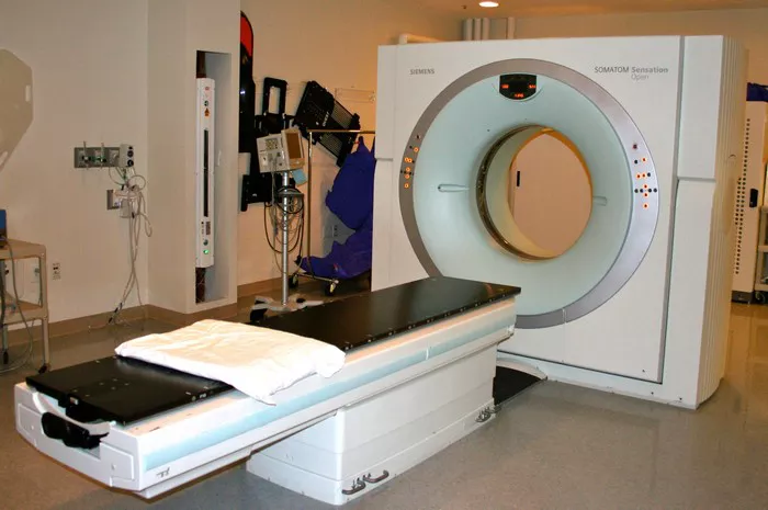
In the world of medical diagnostics, the term CT scanner has become synonymous with detailed, high-quality imaging. But, what exactly is a CT scanner, and how does it work? The CT (computed tomography) scanner is a vital tool in the medical field, providing critical insights into the human body. From emergency rooms to research labs, the CT scanner has revolutionized the way healthcare professionals view internal structures, allowing for faster, more accurate diagnoses.
In this article, we will explore the working principles, components, types, applications, and advancements of CT scanners. Additionally, we will discuss the benefits and potential risks associated with their use.
What is a CT Scanner?
A CT scanner, also known as a CAT scanner (computerized axial tomography), is an advanced medical imaging device that uses X-rays and computer processing to create detailed cross-sectional images, or “slices,” of internal structures within the body. These images are far more detailed than those produced by traditional X-ray imaging, making CT scans a crucial tool for diagnosing various medical conditions.
CT scanners are primarily used in hospitals and clinics, but their applications are also seen in emergency medicine, oncology, orthopedics, neurology, and cardiology, among other fields.
Working Principle of a CT Scanner
The working principle of a CT scanner revolves around the use of X-ray technology combined with powerful computer algorithms to generate detailed images of the body’s internal structures. The process can be broken down into several key steps:
X-ray Generation: At the heart of a CT scanner is an X-ray tube, which generates X-rays. The X-ray tube is mounted on a rotating gantry, a circular frame that allows the tube to rotate around the patient.
Patient Positioning: The patient lies on a motorized table that moves through the center of the scanner. The area of interest is positioned in the center of the scanner’s circular opening, called the gantry.
X-ray Emission and Detection: As the X-ray tube rotates around the patient, it emits X-rays in a fan-like shape. The X-rays pass through the body and are detected by a series of detectors positioned opposite the X-ray tube. These detectors measure the amount of X-rays that pass through the body at different angles.
Image Reconstruction: The detectors send the data to a computer, which reconstructs it into a detailed cross-sectional image of the body. This process involves sophisticated algorithms, such as filtered back projection or iterative reconstruction, which enhance image quality and reduce noise.
Slice Creation: The final output is a series of cross-sectional images (slices) that provide detailed information about the body’s internal structures. These slices can be stacked together to create a 3D representation of the area being scanned.
Key Components of a CT Scanner
A CT scanner is made up of several components, each contributing to the scanning process. The main components include:
X-ray Tube: The X-ray tube generates the X-rays needed for imaging. It emits radiation in multiple directions during rotation.
Detectors: These sensors capture the X-rays that pass through the body. They convert the X-ray data into electrical signals, which are sent to the computer for image reconstruction.
Gantry: The gantry is the circular structure that houses the X-ray tube and detectors. It rotates around the patient during the scan.
Patient Table: This motorized table supports the patient and moves them through the scanner’s gantry. It can be adjusted to target specific areas of the body for imaging.
Computer System: The computer system processes the raw data from the detectors and reconstructs it into detailed images using sophisticated algorithms.
Control Panel: The control panel allows the radiologic technologist or operator to input scan parameters, monitor the scanning process, and access the resulting images.
Types of CT Scanners
CT technology has evolved over time, and various types of CT scanners are now available, each suited for different applications. Some of the major types of CT scanners include:
Single-slice CT Scanners: Early-generation CT scanners that provide one slice per rotation of the gantry. While they offer acceptable image quality, they are slower and less efficient than modern multi-slice scanners.
Multi-slice CT Scanners (Multi-detector CT): These scanners have multiple rows of detectors, allowing for multiple slices to be captured during a single rotation. Multi-slice CT scanners can produce high-resolution images faster than single-slice scanners and are commonly used in modern medical practice.
Spiral (Helical) CT Scanners: These scanners capture continuous slices in a spiral motion as the patient table moves through the gantry. This allows for faster scanning times and better image quality, especially for large areas like the chest or abdomen.
Cone-beam CT: Cone-beam CT scanners are specialized devices used primarily in dental and orthopedic imaging. Unlike traditional CT scanners, which use a fan-shaped beam, cone-beam CT uses a cone-shaped beam to capture images.
Dual-energy CT: A newer type of CT scanner that uses two different X-ray energy levels to create more detailed images. This can help in detecting certain types of tissue abnormalities, such as vascular conditions and tumors.
Applications of CT Scanners
CT scanners have revolutionized the diagnosis and treatment of numerous medical conditions. Some of the most common applications include:
1. Trauma and Emergency Medicine
CT scans are frequently used in emergency settings to rapidly assess injuries. They help identify conditions such as:
- Internal bleeding (e.g., brain hemorrhage)
- Fractures and bone injuries
- Organ damage (e.g., liver or kidney lacerations)
- Trauma to the chest and abdomen
2. Cancer Diagnosis and Treatment
CT scans play a pivotal role in the diagnosis and management of cancers. They are used to:
Detect tumors: CT can help identify the size, location, and extent of tumors.
Monitor tumor growth: Physicians can track the progress of treatment and check for recurrence after surgery or therapy.
Plan radiation therapy: CT scans help in accurately planning radiation treatment by mapping out the tumor’s location.
3. Neurology
CT scanners are commonly used to diagnose neurological conditions, including:
Brain hemorrhages and stroke: Rapid imaging is crucial in these cases to assess bleeding or blockages.
Intracranial pressure: CT scans can provide information about swelling or abnormal pressure within the skull.
Neurological diseases: Conditions such as multiple sclerosis or brain tumors can be detected with a CT scan.
4. Cardiology
CT imaging is also important in cardiology for:
Coronary artery disease: CT angiography allows for the visualization of blood vessels in the heart to detect blockages or narrowing.
Cardiac anatomy: CT scans can evaluate heart structure and function, aiding in the diagnosis of congenital heart defects or valvular diseases.
5. Orthopedics
Orthopedic applications of CT scans include:
Joint and bone injuries: Detailed images help assess fractures, bone infections, or joint dislocations.
Pre-surgical planning: CT scans can be used for planning complex surgeries, such as hip or knee replacements.
6. Abdominal Imaging
CT scans are commonly used to evaluate the abdomen, including conditions such as:
- Appendicitis
- Gallstones
- Liver disease
- Kidney stones
Advantages of CT Scanners
CT scanning has several advantages over traditional X-ray imaging and other diagnostic methods. These include:
High-Resolution Images: CT provides highly detailed images of soft tissues, bones, and organs that are difficult to see with standard X-rays.
Quick and Non-invasive: The scanning process is relatively quick, typically taking just a few minutes. It is also non-invasive, meaning that it does not require surgery or tissue removal to obtain diagnostic information.
3D Imaging: CT scans can generate 3D images from multiple 2D slices, offering a more comprehensive view of the area being studied.
Wide Range of Applications: CT scans can be used for diagnosing a wide variety of conditions across different medical fields.
Guidance for Treatment: CT scans are often used to guide medical procedures, such as biopsies, drainage of abscesses, or placement of stents.
Potential Risks and Considerations
Despite the many benefits, CT scans do come with some risks and considerations:
Radiation Exposure: CT scans involve exposure to X-rays, which can increase the risk of radiation-related health issues, such as cancer. However, the benefits often outweigh the risks, and modern CT scanners have advanced features that minimize radiation doses.
Allergic Reactions to Contrast Agents: Some CT scans require the use of contrast materials to enhance image quality. These agents can sometimes cause allergic reactions or kidney problems in certain individuals.
Limited Soft Tissue Contrast: While CT scans provide excellent detail for bones and some soft tissues, they are less effective than MRI (magnetic resonance imaging) for imaging other types of soft tissues, such as the brain or muscles.
Conclusion
CT scanners are an essential tool in modern medicine, providing detailed, accurate, and fast imaging that aids in the diagnosis, treatment planning, and monitoring of a wide range of medical conditions. Through continuous advancements in technology, CT scanners have become more efficient, safer, and capable of producing even more detailed images with reduced radiation exposure.
As a result, CT scans have proven to be invaluable in fields ranging from emergency medicine to oncology. However, like all medical procedures, their use must be carefully considered to ensure that the benefits outweigh the risks. With ongoing research and development, CT scanners will undoubtedly continue to evolve, improving patient care and outcomes across the globe.

