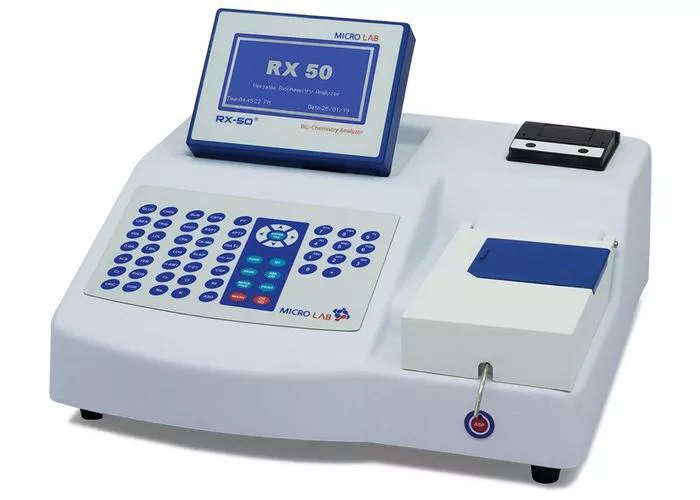
A hematology analyzer is a specialized medical device used to count and analyze the various components of blood. Hematology analyzers provide critical information to healthcare professionals by performing tests that help diagnose and monitor blood-related disorders, such as anemia, leukemia, infections, and other hematological conditions. These analyzers provide results quickly, efficiently, and with high precision, making them an indispensable tool in modern medical laboratories.
In this article, we will explore the working principles of hematology analyzers, including the basic components, types of analysis, and how they use advanced technologies like light scattering, electrical impedance, and fluorescence to perform blood tests. We will also discuss the importance of these machines in clinical diagnostics and their impact on healthcare.
What Is A Hematology Analyzer
Hematology analyzers are designed to automate the process of blood cell analysis, which traditionally involved manual counting and examination under a microscope. The primary function of a hematology analyzer is to count the different types of blood cells—red blood cells (RBCs), white blood cells (WBCs), and platelets—and to measure other critical blood parameters such as hemoglobin concentration, hematocrit (the percentage of blood volume occupied by red blood cells), and mean corpuscular volume (MCV).
Modern hematology analyzers are sophisticated instruments that can provide a wide range of data from a single blood sample. They utilize various principles, including electrical impedance, light scattering, and fluorescence, to analyze blood cells. These principles allow the analyzer to determine cell counts, sizes, and even cell morphology in some cases.
Types of Hematology Analyzers
Hematology analyzers can be broadly categorized into two types: Manual Hematology Analyzers and Automated Hematology Analyzers.
Manual Hematology Analyzers: Manual analyzers, often used in smaller or low-resource settings, typically require human intervention for various stages of the analysis process. These include slide preparation, staining, and microscopic evaluation. The results depend heavily on the expertise of the technician conducting the analysis.
Automated Hematology Analyzers: Automated hematology analyzers have revolutionized blood analysis by providing faster and more accurate results with minimal human intervention. These analyzers are equipped with sensors and software that can automatically perform the entire blood analysis, from sample processing to result interpretation. Modern automated analyzers are capable of producing complete blood counts (CBCs) and other relevant data, such as differential counts and reticulocyte counts, within minutes.
Automated analyzers can further be divided into two categories:
Classic 3-Part Analyzers: These analyzers measure only the three main blood cell types—RBCs, WBCs, and platelets—and provide basic information on the blood composition.
Advanced 5-Part or 6-Part Analyzers: These analyzers not only measure the three main blood components but also categorize different subtypes of white blood cells (e.g., neutrophils, lymphocytes, monocytes, eosinophils, and basophils). They are equipped with additional channels to provide more detailed analysis.
Key Components of a Hematology Analyzer
Regardless of the type, a hematology analyzer typically includes several key components that enable it to perform its analysis effectively. These components include:
Sample Holder: This is where the blood sample is placed for testing. In most modern analyzers, blood is drawn from a patient using an anticoagulant-treated tube. The sample is then processed and transported to the analyzer for testing.
Dilution System: Blood is often diluted to prevent clogging of the analyzer and to achieve optimal flow rates for accurate measurements. The dilution system ensures that the blood sample is mixed with a reagent or diluent to reduce viscosity and maintain sample integrity.
Measurement Chamber: The measurement chamber is where the blood sample is passed through, and various sensors are used to perform the analysis. In many analyzers, the sample is introduced into a flow cytometry chamber or a flow tube, where cells are either passed individually through a sensor or exposed to light or electrical fields.
Electrodes/Sensors: Hematology analyzers use various sensors to detect and measure different parameters of the blood cells. Some analyzers rely on electrical impedance to measure cell volume, while others may use light scattering to determine cell size and complexity.
Optical System: For advanced analyzers, the optical system plays a key role in determining the size, shape, and complexity of blood cells. By passing light through a blood sample or scattering light from individual cells, the analyzer can determine characteristics such as cell morphology, size, and internal structures.
Data Processing Unit: The heart of any automated hematology analyzer is the data processing unit, which interprets the signals received from the sensors and converts them into readable data. This data can be presented in various forms, such as graphs, tables, or numerical values, depending on the analyzer’s design and software.
Display and Interface: The results of the analysis are shown on the display screen of the hematology analyzer. The user interface typically allows technicians to enter patient information, run tests, and interpret results.
How Do Hematology Analyzers Work
The operation of a hematology analyzer involves several sophisticated technologies that work together to provide accurate and reliable results. The most common methods used in automated analyzers include:
1. Electrical Impedance (Coulter Principle)
The Coulter Principle is one of the most widely used methods in hematology analyzers. It involves the measurement of changes in electrical impedance as blood cells pass through a small aperture. When an electrical current flows through the aperture, cells in the sample will cause a disruption in the current, resulting in a voltage pulse. The size of the pulse correlates with the size of the blood cell.
In the Coulter counter, blood is diluted and passed through an orifice that is immersed in an electrolyte solution. As cells pass through the orifice, they displace a volume of electrolyte solution, causing a change in resistance. This change in resistance is detected by the analyzer’s electrodes, and the resulting pulse is used to determine the size of the blood cells.
Red Blood Cells (RBCs): These cells are typically larger than platelets, so their pulse is of a different magnitude.
Platelets: These are smaller and will generate smaller pulses than RBCs.
White Blood Cells (WBCs): These cells are larger than platelets but smaller than RBCs.
This method is particularly effective for counting cells and measuring their volume, making it ideal for performing a complete blood count (CBC).
2. Optical Light Scattering (Laser Flow Cytometry)
Optical light scattering is often used in conjunction with the Coulter Principle to enhance the analysis of blood samples, especially when differentiating between the different types of white blood cells. In this method, the blood sample is exposed to a laser beam as it flows through the analyzer.
When the light hits a cell, it is scattered in different directions depending on the cell’s size, shape, and internal structure. The forward scatter is typically proportional to the cell’s size, while the side scatter can provide information about the granularity and internal complexity of the cell.
For example:
RBCs: These cells have a simple shape and little internal structure, resulting in low light scattering.
WBCs: These cells have a more complex internal structure, leading to more scattered light and allowing for the differentiation of the various subtypes of WBCs, such as neutrophils, lymphocytes, monocytes, eosinophils, and basophils.
This technique is particularly useful for creating detailed white blood cell differential counts and for identifying abnormal cells in blood disorders such as leukemia.
3. Fluorescence Detection
Some advanced hematology analyzers utilize fluorescence to measure specific cellular characteristics, such as DNA or RNA content. This is achieved by staining blood cells with fluorescent dyes that bind to specific cell components.
For instance:
Nucleic Acid Staining: Fluorescent dyes that bind to DNA or RNA can be used to identify and quantify the amount of genetic material in a cell. This is especially helpful in distinguishing between different types of WBCs, as the DNA content varies among different cell types.
Reticulocyte Counting: Reticulocytes are immature red blood cells that are still synthesizing hemoglobin. By staining these cells with a fluorescent dye, the analyzer can identify and count them, providing important information about bone marrow activity and anemia.
Fluorescence detection offers a high degree of specificity and sensitivity, enabling more detailed and accurate blood analysis.
Types of Blood Parameters Measured
Hematology analyzers measure a wide range of parameters, with some of the most common being:
Red Blood Cell Count (RBC): This measures the number of RBCs in a given volume of blood. Low RBC counts can indicate anemia, while high counts may suggest dehydration or polycythemia.
White Blood Cell Count (WBC): This measures the number of WBCs, which are critical for the immune response. Abnormal WBC counts can indicate infections, leukemia, or immune disorders.
Platelet Count: Platelets are essential for blood clotting, and abnormal counts can lead to bleeding disorders or clotting risks.
Hemoglobin (Hb): This measures the concentration of hemoglobin, the protein in RBCs that carries oxygen. Abnormal hemoglobin levels can point to anemia or other blood disorders.
Hematocrit (Hct): This measures the proportion of blood volume that is made up of RBCs.
Mean Corpuscular Volume (MCV): This measures the average size of RBCs. Abnormal MCV values can indicate various types of anemia.
Differential White Blood Cell Count: This breaks down the WBC count into different subtypes, providing important diagnostic information.
Conclusion
Hematology analyzers are complex, high-tech devices that have transformed the field of hematology and clinical diagnostics. By using principles like electrical impedance, optical light scattering, and fluorescence, these machines can quickly and accurately analyze blood samples, providing valuable information to healthcare providers.
The widespread use of automated hematology analyzers has drastically improved diagnostic efficiency, allowing for early detection of blood disorders and better patient management. As technology continues to evolve, we can expect even more precise and comprehensive analysis of blood samples, helping clinicians diagnose and treat a wide range of blood-related conditions more effectively.

